ホーム 計算ツール
代理店ログイン

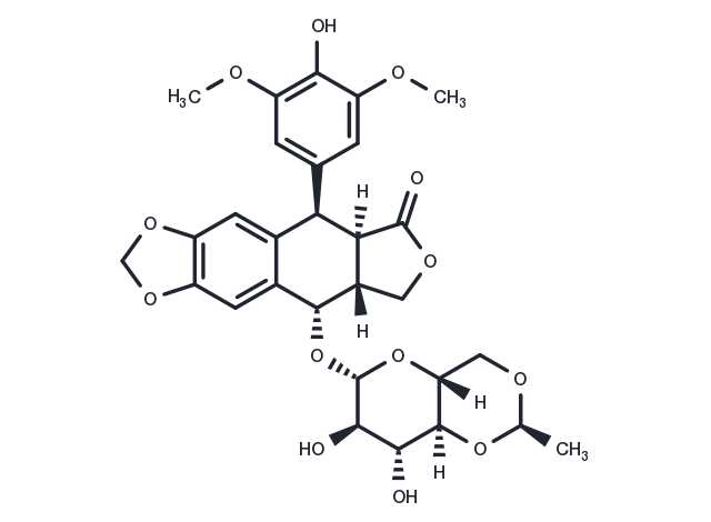
Etoposide

カタログ番号 T0132   CAS 33419-42-0
別名: VP-16, VP-16-213

Etoposide (VP-16-213) is a topoisomerase II inhibitor that inhibits DNA synthesis by forming a complex with topoisomerase II and DNA (IC50=60.3 μM). Etoposide has antitumor activity and induces apoptosis and autophagy.
TargetMolの製品は全て研究用試薬です。人体にはご使用できません。 また、個人の方への販売は行っておりません。
Etoposide, CAS 33419-42-0
パッケージサイズ 在庫状況 単価(税別)
サンプルについてお問い合わせ
50 mg 在庫あり ¥ 7,500
100 mg 在庫あり ¥ 11,000
500 mg 在庫あり ¥ 25,000
1 mL * 10 mM (in DMSO) 在庫あり ¥ 11,500
ご確認事項

1. 1研究室・グループあたり最大5製品までお申し込みいただけます。 同一製品は1回のみとなります。

2. 1回につき最大2製品までのお申し込みが可能です。

3. 2回目以降をご希望の際は、前回ご提供のサンプルの実験結果をオンラインでご提供いただく必要がございます。

4. 2023 年 1 月 20 日より前にサンプルをお申し込みいただいたお客様は、2023 年の無料申請枠にはカウントされませんが、以前の実験結果をご提供いただく必要があります。

Get quote
バッチを選択  
バッチの詳細情報はお問い合わせください
生物学的特性に関する説明
化学的特性
保存条件 & 溶解度情報
説明 Etoposide (VP-16-213) is a topoisomerase II inhibitor that inhibits DNA synthesis by forming a complex with topoisomerase II and DNA (IC50=60.3 μM). Etoposide has antitumor activity and induces apoptosis and autophagy.
ターゲット&IC50 Topo II:60.3 μM
In vitro METHODS: Human cervical cancer cells HeLa were treated with Etoposide (25-400 μM) for 24-48 h, and cell viability was measured by MTT.
RESULTS: Etoposide inhibited the proliferation of Hela cells with IC50s of 167.3 μM and 52.7 μM for 24 h and 48 h, respectively.
The IC50 of Etoposide was 167.3 μM and 52.7 μM at 24 h and 48 h, respectively. [1]
METHODS: Human lung adenocarcinoma cells A549 were treated with Etoposide (0.75-3 μM) for 4 h. The cell cycle was detected by Flow Cytometry.
RESULTS: Etoposide caused a significant decrease in the percentage of A549 cells in G0/G1 and S phases. Meanwhile, the percentage of A549 cells in G2/M phase was significantly increased. [2]
METHODS: Mouse embryonic fibroblast MEFs were treated with Etoposide (1.5-150 μM) for 3-18 h, and the expression levels of target proteins were detected by Western Blot.
RESULTS: 150 μM Etoposide induced a strong cleavage of caspase-3 within 6 h, while 1.5 or 15 μM activated caspase-3 only after 18 h. [3]
In vivo METHODS: To detect anti-tumor activity in vivo, Etoposide (10 mg/kg) and Cisplatin (5-7.5 mg/kg) were intraperitoneally injected every two days for two weeks into KSN nude mice harboring human endometrial adenocarcinoma tumors Ishikawa.
RESULTS: Etoposide, as a single agent, had little or no inhibitory effect on tumor growth, while the combination of Etoposide and Cisplatin significantly inhibited tumor growth. [4]
METHODS: To detect anti-tumor activity in vivo, Etoposide (80 mg/kg in 0.5% methylcellulose) was administered by gavage to immunodeficient mice harboring human glioblastoma tumor U87 once a day for 40 days.
RESULTS: 80 mg/kg Etoposide inhibited U87 tumor growth by 95%. [5]
キナーゼ試験 Nuclear extracts are prepared, and nuclei are isolated. The activity of topoisomerase II is calculated from the percentage of decatenation obtained. Tritiated kinoplast DNA (KDNA 0.22 μg) is used as a substrate. Etoposide and topoisomerase II are incubated for 30 min at 37 ℃ and are stopped with 1% sodium dodecyl sulfate (SDS) and proteinase K (100 μg/mL). The percentages of decatenation and inhibition of topoisomerase II by Etoposide are obtained [5].
細胞研究 After the Etoposide treatment, cells are removed from the dish with phosphate-buffered saline (PBS) containing 0.03% trypsin and 0.27 mM ethylenediaminetetraacetic acid (EDTA) and are diluted into culture dishes in appropriate numbers to yield between 20 and 200 colonies. After 12 days, cultures are fixed with methanol-acetic acid, stained with crystal violet, and scored for colonies containing more than 50 cells [5].
動物実験 The in vivo model for nude mice HB (NMHB) has been established. Only HB cells with embryonal components are grafted and reproduced successfully in this model. Each NMHB subsequently is transplanted into 50 mice for treatment groups. Treatment is initiated when the majority of the tumors reach a volume of 50-100 mm3. The mice are stratified according to their tumor volume and randomly assigned to groups of ten animals each. The animals injected with tumor are given ifosfamide, cisplatin, doxorubicin, etoposide (10 mg/kg/day, i.v.), and carboplatin as single agents in two blocks. One group of ten animals for each original xenograft served as a control group. After initiation of treatment, the tumor growth is recorded at 5-day intervals for 25-30 days and the relative tumor volumes are calculated. Twenty-four hours before the animals are sacrificed, bromodeoxyuridine (BrdU) is injected intraperitoneally for the semiquantitative determination of proliferation activity of the tumor cells (50 μg of BrdU/g body weight) [4].
植物由来
別名 VP-16, VP-16-213
分子量 588.56
分子式 C29H32O13
CAS No. 33419-42-0

保存条件

Powder: -20°C for 3 years | In solvent: -80°C for 1 year

溶解度情報

DMSO: 60 mg/mL (101.94 mM)

参考文献

1. HGN ÇELEBİOĞLU, et al. Effects of thymoquinone and etoposide combination on cell viability and genotoxicity in human cervical cancer hela cells. Istanbul J Pharm. 2022; 52(3):258-264. 2. Litwiniec A, et al. Low-dose etoposide-treatment induces endoreplication and cell death accompanied by cytoskeletal alterations in A549 cells: Does the response involve senescence? The possible role of vimentin. Cancer Cell Int. 2013 Feb 5;13(1):9. 3. Jamil S, et al. Etoposide induces cell death via mitochondrial-dependent actions of p5Cancer Cell Int. 2015 Aug 7;15:79. 4. Suzuki M, et al. Anticancer activity of the combination of cisplatin and etoposide in endometrial cancer-bearing nude mice. Gynecol Oncol. 1991 Apr;41(1):41-5. 5. Panigrahy D, et al. Inhibition of tumor angiogenesis by oral etoposide. Exp Ther Med. 2010 Sep;1(5):739-746. 6. Lee KI, et al. Etoposide induces pancreatic β-cells cytotoxicity via the JNK/ERK/GSK-3 signaling-mediated mitochondria-dependent apoptosis pathway. Toxicol In Vitro. 2016 Jul 26. pii: S0887-2333(16)30147-3. 7. Calvani M, det al. Etoposide-Bevacizumab a new strategy against human melanoma cells expressing stem-like traits. Oncotarget. 2016 Jun 9. doi: 10.18632/oncotarget.9939. 8. Zhang J, Hirst A J, Duan F, et al. Darren Robinson, 3 Mark Jones, 2 Le Li, 4 Peizhe Wang, Peng Jiang, 4 Peter W. Andrews, 2 Ivana Barbaric, 2,* and Jie Na[J]. Anti-apoptotic Mutations Desensitize Human Pluripotent Stem Cells to Mitotic Stress and Enable Aneuploid Cell Survival. Stem Cell Reports. 9. Ruan C, Wang C, Gong X, et al. An integrative multi-omics approach uncovers the regulatory role of CDK7 and CDK4 in autophagy activation induced by silica nanoparticles[J]. Autophagy. 2020. 10. Weizhe Li, Hong-Yan Wang, Xiaolu Zhao, Hongguo Duan, Binghua Cheng, Yafei Liu, Mengjie Zhao et al. A methylation-phosphorylation switch determines Plk1 kinase activity and function in DNA damage repair [J]. Science Advances. 2019 Mar 6;5(3):eaau7566.

引用文献

1. Yang C, Xu H, Yang D, et al.A renal YY1-KIM1-DR5 axis regulates the progression of acute kidney injury.Nature Communications.2023, 14(1): 4261. 2. Qu Y Q, Song L L, Xu S W, et al.Pomiferin targets SERCA, mTOR, and P-gp to induce autophagic cell death in apoptosis-resistant cancer cells, and reverses the MDR phenotype in cisplatin-resistant tumors in vivo.Pharmacological Research.2023: 106769. 3. Wang D, Wang Y, Di X, et al.Cortical tension drug screen links mitotic spindle integrity to Rho pathway.Current Biology.2023 4. Ruan C, Wang C, Gong X, et al. An integrative multi-omics approach uncovers the regulatory role of CDK7 and CDK4 in autophagy activation induced by silica nanoparticles. Autophagy. 2020 5. Yang G, Wan P, Xiang Q, et al. E3 Ubiquitin Ligase ASB17 Promotes Apoptosis by Ubiquitylating and Degrading BCLW and MCL1. Biology-Basel. 2021, 10(3): 234. 6. Feng J, Xi Z, Jiang X, et al. Saikosaponin a enhances Docetaxel efficacy by selectively inducing death of dormant prostate cancer cells through excessive autophagy. Cancer Letters. 2022: 216011. 7. Li W, Wang H Y, Zhao X, et al. A methylation-phosphorylation switch determines Plk1 kinase activity and function in DNA damage repair. Science Advances. 2019, 5(3): eaau7566 8. Zhang J, Hirst A J, Duan F, et al. Darren Robinson, 3 Mark Jones, 2 Le Li, 4 Peizhe Wang, Peng Jiang, 4 Peter W. Andrews, 2 Ivana Barbaric, 2,* and Jie Na[J]. Anti-apoptotic Mutations Desensitize Human Pluripotent Stem Cells to Mitotic Stress and Enable Aneuploid Cell Survival. Stem Cell Reports. 2019, 12(3): 557-571 9. Kong Y, Liu Y, Li X, et al.Palmitoylation landscapes across human cancers reveal a role of palmitoylation in tumorigenesis.Journal of Translational Medicine.2023, 21(1): 1-19. 10. Wu X, Yi X, Zhao B, et al.The volume regulated anion channel VRAC regulates NLRP3 inflammasome by modulating itaconate efflux and mitochondria function.Pharmacological Research.2023: 107016.
隠し

関連化合物ライブラリー

この製品は下記化合物ライブラリに含まれています:
Traditional Chinese Medicine Monomer Library Anti-Cancer Approved Drug Library Drug Repurposing Compound Library Natural Product Library for HTS Anti-Cancer Active Compound Library Anti-Cancer Clinical Compound Library Miao medicine Compound Library Inhibitor Library Anti-Cancer Drug Library Microbial Natural Product Library

関連製品

同一標的の関連化合物
Nerol Isatuximab Acitretin Etidronic acid Stavudine ROC-325 Enzastaurin Abacavir sulfate

投与量変換

You can also refer to dose conversion for different animals. 詳細

In vivo投与量計算 (透明溶液)

ステップ1: 以下の情報を入力してください
投与量
mg/kg
動物の平均体重
g
動物あたりの投与量
ul
動物数
溶媒の組成を入力してください
% DMSO
%
% Tween 80
% ddH2O
計算する リセット

計算器

モル濃度計算機
希釈計算機
再構成計算
分子量計算機
=
X
X

モル度計算機では以下の計算が可能です

  • 既知の体積と濃度の溶液を調製するために必要な化合物の質量
  • 質量が既知の化合物を目的の濃度まで溶解させるのに必要な溶液の量
  • 特定の体積の中に既知の質量の化合物を入れて得られる溶液の濃度
参考例

モル濃度計算機を使用したモル濃度計算の例
化合物の分子量が197.13g/molである場合、10mlの水に10mMのストック溶液を作るのに必要な化合物の質量はどれくらいですか?
[分子量(MW)]の欄に[197.13]と入力してください
[濃度]ボックスに10と入力し、正しい単位(millimolar)を選択します
[容量]ボックスに10と入力し、正しい単位(milliliter)を選択します
計算を押します
答えの19.713mgが質量欄に表示されます

X
=
X

溶液を作るのに必要な希釈率の計算

溶液の調製に必要な希釈率の算出
希釈計算機は、既知の濃度の原液をどのように希釈するかを計算することができる便利なツールです。V1を計算するためにC1、C2&V2を入力します。

参考例

Tocrisの希釈計算器を用いた希釈計算の一例
50μMの溶液を20ml作るためには、10mMの原液を何ml必要ですか?
C1V1=C2V2という式を用いて、C1=10mM、C2=50μM、V2=20ml、V1を未知数とします。
濃度(開始)ボックスに10を入力し正しい単位(millimolar)を選択してください
濃度(終了)ボックスに50を入力し正しい単位(millimolar)を選択してください
体積(終了)ボックスに20を入力し正しい単位(millimolar)を選択してください
計算を押します
100 microliter (0.1 ml) という答えが体積(開始)ボックスに表示されます。

=
/

バイアルを再構成するのに必要な溶媒の量を計算する.

再構成計算機を使えば、バイアルを再構成するための試薬の量をすぐに計算することができます.
試薬の質量と目標濃度を入力するだけで計算します。

g/mol

化合物の化学式を入力して、そのモル質量や元素組成を計算します

Tヒント:化学式は大文字と小文字を区別します。: C10H16N2O2 c10h16n2o2

化合物のモル質量(分子量)を計算する手順:
化学物質のモル質量を計算するには、その化学式を入力し、「計算」をクリックしてください。.
分子質量、分子量、モル質量、モル重量の定義:
分子質量(分子量)とは、物質の1分子の質量であり、統一された原子質量単位(u)で表されます。(1uは炭素12の1原子の質量の1/12に等しい)
モル質量(molar weight)とは、ある物質の1モルの質量のことで、単位はg/molです。

bottom

技術サポート

Please see Inhibitor Handling Instructions for more frequently ask questions. Topics include: how to prepare stock solutions, how to store products, and cautions on cell-based assays & animal experiments, etc.

Keywords

Etoposide 33419-42-0 Apoptosis Autophagy DNA Damage/DNA Repair Microbiology/Virology Mitophagy Antibiotic Topoisomerase Antibacterial VP-16 P388 HCT116 p53 chemotherapy Mitochondrial Autophagy FBXW Inhibitor anti-cancer inhibit VP 16 Bacterial prodrug VP-16-213 VP16 leukemia inhibitor