ホーム 計算ツール
代理店ログイン

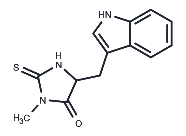
Necrostatin-1

カタログ番号 T1847   CAS 4311-88-0
別名: Nec-1, Necrostatin 1

Necrostatin-1 (Nec-1) is a necrotic apoptosis inhibitor and RIP1 inhibitor with specificity. Necrostatin-1 inhibits TNF-α-induced necrotic apoptosis. Necrostatin-1 also inhibits IDO.
TargetMolの製品は全て研究用試薬です。人体にはご使用できません。 また、個人の方への販売は行っておりません。
Necrostatin-1, CAS 4311-88-0
パッケージサイズ 在庫状況 単価(税別)
サンプルについてお問い合わせ
5 mg 在庫あり ¥ 7,000
10 mg 在庫あり ¥ 10,500
25 mg 在庫あり ¥ 17,500
50 mg 在庫あり ¥ 26,500
100 mg 在庫あり ¥ 41,000
200 mg 在庫あり ¥ 60,500
500 mg 在庫あり ¥ 101,500
1 mL * 10 mM (in DMSO) 在庫あり ¥ 11,000
ご確認事項

1. 1研究室・グループあたり最大5製品までお申し込みいただけます。 同一製品は1回のみとなります。

2. 1回につき最大2製品までのお申し込みが可能です。

3. 2回目以降をご希望の際は、前回ご提供のサンプルの実験結果をオンラインでご提供いただく必要がございます。

4. 2023 年 1 月 20 日より前にサンプルをお申し込みいただいたお客様は、2023 年の無料申請枠にはカウントされませんが、以前の実験結果をご提供いただく必要があります。

Get quote
バッチを選択  
バッチの詳細情報はお問い合わせください
生物学的特性に関する説明
化学的特性
保存条件 & 溶解度情報
説明 Necrostatin-1 (Nec-1) is a necrotic apoptosis inhibitor and RIP1 inhibitor with specificity. Necrostatin-1 inhibits TNF-α-induced necrotic apoptosis. Necrostatin-1 also inhibits IDO.
ターゲット&IC50 RIP1:490 nM(EC50, Jurkat cells)
In vitro METHODS: Human hepatocellular carcinoma cells Huh7 and SK-HEP-1 were pretreated with Necrostatin-1 (10-20 µM) for 1 h, and then treated with sulfasalazine, erastin or RSL3 for 24 h. Cell viability was measured by CellTiter Glo® assay.
RESULTS: Necrostatin-1 significantly blocked the decrease in cell viability induced by sulfasalazine and erastin in both cell lines and partially reversed the decrease in cell viability induced by RSL3 in SK-HEP-1 cells. [1]
METHODS: Human histiocytic lymphoma cells U937 were treated with Necrostatin-1 (1-20 µM), zVAD.fmk (100 μM), and TNFα (10 ng/mL) for 72 h. Cell viability was detected by ATP-based viability assay.
RESULTS: Necrostatin-1 effectively blocked the necrotic death of U937 cells in a concentration-dependent manner. [2]
METHODS: H/R injury-induced human renal papillomatous cells HK-2 were treated with Necrostatin-1 (30 mmol/L) for 2-12 h. Cell death was analyzed by Flow Cytometry.
RESULTS: Necrostatin-1 partially protected HK-2 cells from H/R-induced necrosis. [3]
In vivo METHODS: To study the pathophysiology of contrast-induced AKI (CIAKI), Necrostatin-1 (1.65 mg/kg) was administered intraperitoneally as a single injection to C57BL/6 mice, and CIAKI was induced by using radiocontrast media (RCM) 15 min later.
RESULTS: Necrostatin-1 prevented osmotic nephropathy and CIAKI. Necrostatin-1 blocked RCM-induced peritubular capillary dilatation, suggesting that the structural domain of RIP1 kinase has a novel role in regulating the microvascular hemodynamics and pathophysiology of CIAKI that is independent of cell death. [4]
METHODS: To investigate the protective effect and mechanism of hepatitis in mice, Necrostatin-1 (1.8 mg/kg) was administered intraperitoneally to C57BL/6 mice as a single injection, and concanavalin A was used to induce hepatitis 1 h later.
RESULTS: Improvements in liver function and histopathologic changes, as well as suppression of inflammatory cytokine production, were observed in Necrostatin-1-injected mice. The expression of TNF-α, IFN-γ, IL2, IL6, and RIP1 was significantly reduced in Necrostatin-1-injected mice, and autophagosome formation was significantly reduced by Necrostatin-1 treatment. The RESULTS suggest that Necrostatin-1 prevents concanavalin A-induced liver injury through RIP1-related and autophagy-related pathways. [5]
キナーゼ試験 The assay was performed essentially as described. 293T cells were transfected with pcDNA3-FLAG-RIP1 vector, vectors encoding RIP1 mutant proteins or pcDNA3-RIP2-Myc and pcDNA3-FLAG-RIP3 vectors using standard Ca3(PO4)2 precipitation procedure. Culture medium was replaced 6 h after the transfection and cells were lysed 48 h later in the TL buffer consisting of 1% Triton X-100, 150 mM NaCI, 20 mM HEPES, pH 7.3, 5 mM EDTA, 5 mM NaF, 0.2 mM NaVO3 and complete protease inhibitor cocktail. Immunoprecipitation was carried out for 16 h at 4 °C using anti-FLAG M2 agarose beads, followed by three washes with TL buffer and two washes with 20 mM HEPES, pH 7.3. Beads were incubated in 15 μl of the reaction buffer containing 20 mM HEPES, pH 7.3, 10 mM MnCl2 and 10 mM MgCl2 for 15 min at 23–25 °C in the presence of different concentrations of necrostatins. For these assays, compound stocks (in DMSO) were diluted to appropriate concentrations in DMSO before the addition to the reactions to maintain final concentration of DMSO for all samples at 3%. Kinase reaction was initiated by addition of 10 μM cold ATP and 1 mCi of [γ-32P] ATP, and reactions were carried out for 30 min at 30 °C. Reactions were stopped by boiling in SDS-PAGE sample buffer and subjected to 8% SDS-PAGE. RIP1 band was visualized by analysis in a Storm 8200 Phosphorimager. Similar protocol was used for endogenous RIP1 kinase reactions, except mouse monoclonal RIP1 antibody and protein magnetic beads or rabbit RIP1 antibody-coupled agarose beads were used. For recombinant baculovirally expressed RIP1, protein was expressed in Sf9 cells according to manufacturer's instructions and purified using glutathione-sepharose beads. Protein was eluted in 50 mM Tris-HCl, pH 8.0 supplemented with 10 mM reduced glutathione, and eluted protein was used in the kinase reactions, supplemented with 5 × kinase reaction buffer (100 mM HEPES, pH 7.3, 50 mM MnCl2, 50 mM MgCl2, 50 μM cold ATP and 5 μCi of [γ-32P]ATP) [1].
細胞研究 Determination of EC50 was performed in FADD-deficient Jurkat cells treated with human TNFα as previously described. Briefly, cells were seeded into 96-well plates and treated with a range of necrostatin concentrations (30 nM to 100 μM, 11 dose points) in the presence and absence of 10 ng ml–1 human TNFα for 24 h. For these and all other cellular assays, compound stocks (in DMSO) were diluted to appropriate concentrations in DMSO before addition to the cells to maintain final concentration of DMSO for all samples at 0.5%. Cell viability was determined using CellTiter-Glo luminescent cell viability assay. Ratio of luminescence in compound and TNF-treated wells to compound-treated, TNF-untreated wells was calculated (viability, %) [1].
動物実験 24 hours after reperfusion, mice received intravenous application of 200 μl PBS or RCM via the tail vein. A single dose of zVAD (10 mg/kg body weight) or Nec-1 (1.65 mg/kg body weight) was applied intraperitoneally 15 min. before RCM-injection. To test the activity of zVAD, we applied zVAD from the same byculture to anti-Fas-treated Jurkat cells to assure its quality before mice were treated with this compound. Mice were harvested another 24 hours after RCM-application (48 hours after reperfusion). Blood samples were obtained from retroorbital bleeding and serum levels of urea and creatinine 5 were determined according to clinical standards in the central laboratory of the University Hospital Schleswig-Holstein, Campus Kiel, Germany, employing an enzymatic ultraviolettest for urea and an enzymatic peroxidase-dependent test for creatinine according to the manufacturer's instructions. Kidneys were conserved for histology. In addition to the demonstrated experiments, we compared the PBS group to mice that only received IRI without 200 μl of PBS and detected no changes in serum concentrations of urea and creatinine or histologically [3].
別名 Nec-1, Necrostatin 1
分子量 259.33
分子式 C13H13N3OS
CAS No. 4311-88-0

保存条件

Powder: -20°C for 3 years | In solvent: -80°C for 1 year

溶解度情報

DMSO: 40 mg/mL (154.24 mM)

参考文献

1. Hanna Y, et al. Necrostatin-1 Prevents Ferroptosis in a RIPK1- and IDO-Independent Manner in Hepatocellular Carcinoma. Antioxidants. 2021 July;10(9):1347. 2. Degterev A, et al. Chemical inhibitor of nonapoptotic cell death with therapeutic potential for ischemic brain injury. Nat Chem Biol. 2005 Jul;1(2):112-9. doi: 10.1038/nchembio711. Epub 2005 May 29. Erratum in: Nat Chem Biol. 2005 Sep;1(4):234. 3. Shen B, et al. Necrostatin-1 Attenuates Renal Ischemia and Reperfusion Injury via Meditation of HIF-1α/mir-26a/TRPC6/PARP1 Signaling. Mol Ther Nucleic Acids. 2019 Sep 6;17:701-713. 4. Linkermann A, et al. The RIP1-kinase inhibitor necrostatin-1 prevents osmotic nephrosis and contrast-induced AKI in mice. J Am Soc Nephrol. 2013 Oct;24(10):1545-57. 5. Zhou Y, et al. Protective effects of necrostatin-1 against concanavalin A-induced acute hepatic injury in mice. Mediators Inflamm. 2013;2013:706156. 6. hang C, Liu Z, Zhang Y, et al. Z“Iron free” zinc oxide nanoparticles with ion-leaking properties disrupt intracellular ROS and iron homeostasis to induce ferroptosis[J]. Cell Death & Disease. 2020, 11(3): 1-15. 7. Yao X, Ma S, Peng S, et al. Zwitterionic Polymer Coating of Sulfur Dioxide‐Releasing Nanosystem Augments Tumor Accumulation and Treatment Efficacy[J]. Advanced Healthcare Materials. 2020, 9(5): 1901582. 9. Wang S, Li F, Qiao R, et al. Arginine-Rich Manganese Silicate Nanobubbles as a Ferroptosis-Inducing Agent for Tumor-Targeted Theranostics[J]. ACS nano. 2018 Dec 26;12(12):12380-12392. 10. Yan B, Ai Y, Sun Q, et al. Membrane Damage during Ferroptosis Is Caused by Oxidation of Phospholipids Catalyzed by the Oxidoreductases POR and CYB5R1[J]. Molecular Cell. 2020

引用文献

1. Hu G, Cui Z, Chen X, et al.Suppressing Mesenchymal Stromal Cell Ferroptosis Via Targeting a Metabolism‐Epigenetics Axis Corrects their Poor Retention and Insufficient Healing Benefits in the Injured Liver Milieu.Advanced Science.2023: 2206439. 2. Li Y, Yang W, Zheng Y, et al.Targeting fatty acid synthase modulates sensitivity of hepatocellular carcinoma to sorafenib via ferroptosis.Journal of Experimental & Clinical Cancer Research.2023, 42(1): 1-19. 3. Wang X, Ji Y, Qi J, et al.Mitochondrial carrier 1 (MTCH1) governs ferroptosis by triggering the FoxO1-GPX4 axis-mediated retrograde signaling in cervical cancer cells.Cell Death & Disease.2023, 14(8): 1-13. 4. Lei S, Chen C, Han F, et al.AMER1 deficiency promotes the distant metastasis of colorectal cancer by inhibiting SLC7A11-and FTL-mediated ferroptosis.Cell Reports.2023, 42(9). 5. Zhou R, You Y, Zha Z, et al.Biotin decorated celastrol-loaded ZIF-8 nano-drug delivery system targeted epithelial ovarian cancer therapy.Biomedicine & Pharmacotherapy.2023, 167: 115573. 6. Zhu X, Huang N, Ji Y, et al.Brusatol induces ferroptosis in oesophageal squamous cell carcinoma by repressing GSH synthesis and increasing the labile iron pool via inhibition of the NRF2 pathway.Biomedicine & Pharmacotherapy.2023, 167: 115567. 7. Li H, Guan J, Chen J, et al.Necroptosis signaling and NLRP3 inflammasome cross-talking in epithelium facilitate Pseudomonas aeruginosa mediated lung injury.Biochimica et Biophysica Acta (BBA)-Molecular Basis of Disease.2022: 166613. 8. Wu Z, Lin C, Zhang F, et al.TIGD1 Function as a Potential Cuproptosis Regulator Following a Novel Cuproptosis-Related Gene Risk Signature in Colorectal Cancer.Cancers.2023, 15(8): 2286. 9. Huang F, Liang J, Lin Y, et al.Repurposing of Ibrutinib and Quizartinib as potent inhibitors of necroptosis.Communications Biology.2023, 6(1): 972. 10. Cai H, Qin D, Liu Y, et al.Remodeling of Gut Microbiota by Probiotics Alleviated Heat Stroke‐Induced Necroptosis in Male Germ Cells.Molecular Nutrition & Food Research.2023: 2300291.
11. Zeng H, Xie H, Ma Q, et al.Identification of N-(3-(methyl (3-(orotic amido) propyl) amino) propyl) oleanolamide as a novel topoisomerase I catalytic inhibitor by rational design, molecular dynamics simulation, and biological evaluation.Bioorganic Chemistry.2023: 106734. 12. Du S, Zeng F, Sun H, et al. Prognostic and therapeutic significance of a novel ferroptosis related signature in colorectal cancer patients. Bioengineered. 2022, 13(2): 2498-2512. 13. Ning X, Qi H, Yuan Y, et al. Identification of a new small molecule that initiates ferroptosis in cancer cells by inhibiting the system Xc− to deplete GSH. European Journal of Pharmacology. 2022: 175304. 14. Wang S, Li F, Qiao R, et al. Arginine-Rich Manganese Silicate Nanobubbles as a Ferroptosis-Inducing Agent for Tumor-Targeted Theranostics. ACS nano. 2018 Dec 26;12(12):12380-12392. 15. Su G, Yang W, Wang S, et al. SIRT1-autophagy axis inhibits excess iron-induced ferroptosis of foam cells and subsequently increases IL-1Β and IL-18. Biochemical and Biophysical Research Communications. 2021, 561: 33-39. 16. Wu X, Lu Y, Qin X. Combination of Compound Kushen Injection and cisplatin shows synergistic antitumor activity in p53-R273H/P309S mutant colorectal cancer cells through inducing apoptosis. Journal of Ethnopharmacology. 2021: 114690. 17. Yao X, Ma S, Peng S, et al. Zwitterionic Polymer Coating of Sulfur Dioxide‐Releasing Nanosystem Augments Tumor Accumulation and Treatment Efficacy. Advanced Healthcare Materials. 2020, 9(5): 1901582. 18. Wang F, Xie M, Chen P, et al. Homoharringtonine combined with cladribine and aclarubicin (HCA) in acute myeloid leukemia: A new regimen of conventional drugs and its mechanism. Oxidative Medicine and Cellular Longevity. 2022 19. Yang W, Liu S, Li Y, et al. Pyridoxine induces monocyte-macrophages death as specific treatment of acute myeloid leukemia. Cancer Letters. 2020 20. Ni H, Qin H, Sun C, et al. MiR-375 reduces the stemness of gastric cancer cells through triggering ferroptosis. Stem Cell Research & Therapy. 2021, 12(1): 1-17. 21. Zhang Y, Fan B Y, Pang Y L, et al. Neuroprotective effect of deferoxamine on erastininduced ferroptosis in primary cortical neurons. Neural Regeneration Research. 2020, 15(8): 1539 22. Yan B, Ai Y, Sun Q, et al. Membrane Damage during Ferroptosis Is Caused by Oxidation of Phospholipids Catalyzed by the Oxidoreductases POR and CYB5R1. Molecular Cell. 2020 23. hang C, Liu Z, Zhang Y, et al. Z“Iron free” zinc oxide nanoparticles with ion-leaking properties disrupt intracellular ROS and iron homeostasis to induce ferroptosis. Cell Death & Disease. 2020, 11(3): 1-15. 24. Yang K H, Tang J Y, Chen Y N, et al. Nepenthes Extract Induces Selective Killing, Necrosis, and Apoptosis in Oral Cancer Cells. Journal of Personalized Medicine. 2021, 11(9): 871. 25. D’Onofrio N, Martino E, Balestrieri A, et al. Diet‐derived ergothioneine induces necroptosis in colorectal cancer cells by activating the SIRT3/MLKL pathway. FEBS letters. 2022 26. Wu H, Cheng X, Huang F, et al. Aprepitant Sensitizes Acute Myeloid Leukemia Cells to the Cytotoxic Effects of Cytosine Arabinoside in vitro and in vivo. Development and Therapy. 2020, 14: 2413 27. Wang Z, Zou F, Wang A, et al. Repurposing of the FGFR inhibitor AZD4547 as a potent inhibitor of necroptosis by selectively targeting RIPK1. Acta Pharmacologica Sinica. 2022: 1-10 28. Wang Y, Zhang B, Liu S, et al.The traditional herb Sargentodoxa cuneata alleviates DSS-induced colitis by attenuating epithelial barrier damage via blocking necroptotic signaling.Journal of Ethnopharmacology.2023: 117373. 29. Chen H, Hu J, Xiong X, et al.AURKA inhibition induces Ewing’s sarcoma apoptosis and ferroptosis through NPM1/YAP1 axis.Cell Death & Disease.2024, 15(1): 99. 30. Li J, Liu X, Liu Y, et al.Saracatinib inhibits necroptosis and ameliorates psoriatic inflammation by targeting MLKL.Cell Death & Disease.2024, 15(2): 122. 31. Chen J, Liu Y, You Y, et al.Biotin-decorated celastrol-loaded ZIF-8 nanoparticles induce ferroptosis for colorectal cancer therapy.Materials & Design.2024: 112814.
隠し

関連化合物ライブラリー

この製品は下記化合物ライブラリに含まれています:
Inhibitor Library Kinase Inhibitor Library Antidepressant Compound Library Bioactive Compounds Library Max Anti-Liver Cancer Compound Library Anti-Ovarian Cancer Compound Library Bioactive Compound Library NF-κB Signaling Compound Library NO PAINS Compound Library Anti-Cancer Metabolism Compound Library

関連製品

同一標的の関連化合物
IDO-IN-7 Coptisine 4-Phenyl-1H-1,2,3-triazole IDO inhibitor 1 Palmatine IDO1-IN-21 IDO1/TDO-IN-6 Indoximod

投与量変換

You can also refer to dose conversion for different animals. 詳細

In vivo投与量計算 (透明溶液)

ステップ1: 以下の情報を入力してください
投与量
mg/kg
動物の平均体重
g
動物あたりの投与量
ul
動物数
溶媒の組成を入力してください
% DMSO
%
% Tween 80
% ddH2O
計算する リセット

計算器

モル濃度計算機
希釈計算機
再構成計算
分子量計算機
=
X
X

モル度計算機では以下の計算が可能です

  • 既知の体積と濃度の溶液を調製するために必要な化合物の質量
  • 質量が既知の化合物を目的の濃度まで溶解させるのに必要な溶液の量
  • 特定の体積の中に既知の質量の化合物を入れて得られる溶液の濃度
参考例

モル濃度計算機を使用したモル濃度計算の例
化合物の分子量が197.13g/molである場合、10mlの水に10mMのストック溶液を作るのに必要な化合物の質量はどれくらいですか?
[分子量(MW)]の欄に[197.13]と入力してください
[濃度]ボックスに10と入力し、正しい単位(millimolar)を選択します
[容量]ボックスに10と入力し、正しい単位(milliliter)を選択します
計算を押します
答えの19.713mgが質量欄に表示されます

X
=
X

溶液を作るのに必要な希釈率の計算

溶液の調製に必要な希釈率の算出
希釈計算機は、既知の濃度の原液をどのように希釈するかを計算することができる便利なツールです。V1を計算するためにC1、C2&V2を入力します。

参考例

Tocrisの希釈計算器を用いた希釈計算の一例
50μMの溶液を20ml作るためには、10mMの原液を何ml必要ですか?
C1V1=C2V2という式を用いて、C1=10mM、C2=50μM、V2=20ml、V1を未知数とします。
濃度(開始)ボックスに10を入力し正しい単位(millimolar)を選択してください
濃度(終了)ボックスに50を入力し正しい単位(millimolar)を選択してください
体積(終了)ボックスに20を入力し正しい単位(millimolar)を選択してください
計算を押します
100 microliter (0.1 ml) という答えが体積(開始)ボックスに表示されます。

=
/

バイアルを再構成するのに必要な溶媒の量を計算する.

再構成計算機を使えば、バイアルを再構成するための試薬の量をすぐに計算することができます.
試薬の質量と目標濃度を入力するだけで計算します。

g/mol

化合物の化学式を入力して、そのモル質量や元素組成を計算します

Tヒント:化学式は大文字と小文字を区別します。: C10H16N2O2 c10h16n2o2

化合物のモル質量(分子量)を計算する手順:
化学物質のモル質量を計算するには、その化学式を入力し、「計算」をクリックしてください。.
分子質量、分子量、モル質量、モル重量の定義:
分子質量(分子量)とは、物質の1分子の質量であり、統一された原子質量単位(u)で表されます。(1uは炭素12の1原子の質量の1/12に等しい)
モル質量(molar weight)とは、ある物質の1モルの質量のことで、単位はg/molです。

bottom

技術サポート

Please see Inhibitor Handling Instructions for more frequently ask questions. Topics include: how to prepare stock solutions, how to store products, and cautions on cell-based assays & animal experiments, etc.

Keywords

Necrostatin-1 4311-88-0 Apoptosis Autophagy Metabolism NF-Κb Indoleamine 2,3-Dioxygenase (IDO) Ferroptosis RIP kinase Receptor-interacting protein kinases Inhibitor RIPK Nec 1 Necrostatin1 Nec1 Nec-1 inhibit Necrostatin 1 inhibitor